Research topics of the Brain and Perception axis
Many neuroimaging techniques such as visual event-related potentials, magnetoencephalography, or even magnetic resonance imaging allow a better understanding of the visual functions in humans and animal models. The Perception and Brain axis builds on new technological breakthroughs in imaging based on its important contribution to the comprehension of mechanisms underlying normal and pathologic visual processes.
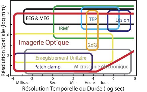
Source: Grinvald 2005
The examples of projects described below use these modern techniques, to which are added complementary electrophysiological, psychophysical, pharmacological, behavioural, neuroanatomical and biomedical approaches.
Neurophysiological correlates of visual perception
Considering the importance of movement in everyday life, many members of the axis study the nervous pathways and the structures responsible for the analysis of global movement. For example, the analysis of the optical flow created during locomotion, implying the integration of local indexes, is essential to the normal visuomotor behaviour and, consequently, to autonomous locomotion. A particular attention is paid to the linear and non-linear mechanisms involved in the perception of motion. Recording and cell stimulation studies seek to understand the nature of neural coding in different cortical and subcortical visual areas. Other research projects are carried out to study visual functions (e.g. functional integrity, ventral and dorsal pathways, binocular vision, etc.) in normal individuals or in those suffering from pathologies such as autism, prematurity or amblyopia.

 A
A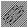
B
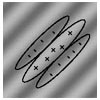
A) Optimal visual stimulus (of the first order) for the encoding of receptive fields in a linear model. B) Stimulus (of the second order), which cannot be decoded by a linear receptive field, requiring non-linear detectors. This type of stimuli is used in many studies.
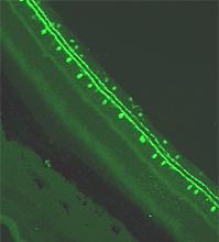
The visual system can be studied by neuroanatomical and electrophysiological techniques. This photo shows a dopaminergic amacrine cell in the rat retina. Source: Christian Casanova’s Laboratory.
The Brain and Perception axis researchers use cutting-edge technology, such as trans-cranial stimulation, optic imagery and, more recently, optogenetics. This latter method associates optics and genetics in order to specifically stimulate one cell type or neuronal group. Using targeted light stimulation, it is possible to map certain neuronal networks among others, opening new fundamental and clinical research avenues. In the animal model, it has been demonstrated that optogenetics allows the reactivation of retinal photosensitivity despite the lack of functional photoreceptors. Indeed, after the integration of photosensitive pigments coupled to ionophorus in retinal cells, a light beam can activate labeled cells and thus allow the transmission of their neural message to the cortex via the optic nerve.
Neuronal plasticity, sensory substitution and residual vision
Following trauma or injury, the visual brain can reorganize and allow the affected individual to recover certain sensory functions. Studies conducted on brain plasticity aim to determine the nature of neurons and visual pathways that resist injury and the various factors responsible for neuronal survival in the injured brain. For example, many researchers are interested in investigating the integrity of retinal ganglion cells after optic nerve or primary visual cortex lesion. The neuroanatomical approach will allow the determination of the effect of neurotrophins on retinal neuron survival rate, their cell type and the nature of surviving retinofugal projections. In parallel, the electrophysiological approach will allow us to determine the functional status of these neurons and the pathways they are associated with. Through these studies, we will be able to identify the factors that may explain or facilitate the establishment of new brain connections.
Furthermore, members of the axis continue to work on the mechanisms underlying sensory substitution. For example, young animals whose visual cortex was injured at birth, thus theoretically blind, can still visually find their way in a labyrinth in adulthood. A selective surgical manipulation allowed the reorientation of the neural circuits starting at the retina to the auditory cortex! Other human studies have shown that blind people have better perceptual abilities, including the localization of sounds in space. It is likely that the visual cortex lacking visual afferent pathways plays a role in this task. Thus, it is now clear that the brain areas that do not receive their normal messages are able to reorganize and participate in monitoring other sensory functions. These studies will allow us to promote the recruitment of cortical regions, wrongly assumed inactive, to improve other brain functions.
Clinical applications of sensory substitution have emerged during the past few years. For example, it is possible to substitute vision through neurostimulators placed on the tongue. Due to neural plasticity, signals from peripheral receptors can relay an equivalent stimulation to the visual cortex. It has been also shown that visual sensations may be generated by sound stimulation.
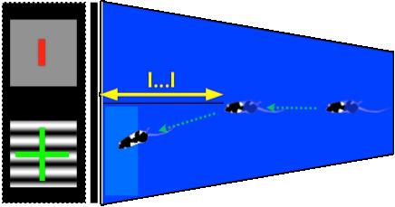
Rodents performing a visual discrimination test. Source: www.cerebralmechanics.com
The members of the axis are also interested in the evaluation of the residual functions in patients with cerebral lesions. Psycho-physical approaches are used to determine the nature of the unaffected visual functions depending on the age of the patient, the extent and the location of the cortical lesions. Studies based on brain imaging are performed to determine the neural pathways involved in the preservation of visual functions and the regions responsible of eye movements associated with residual vision, as well as to document the oculomotor disorders associated with certain therapeutic lesions (eg. epileptic patients).
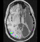
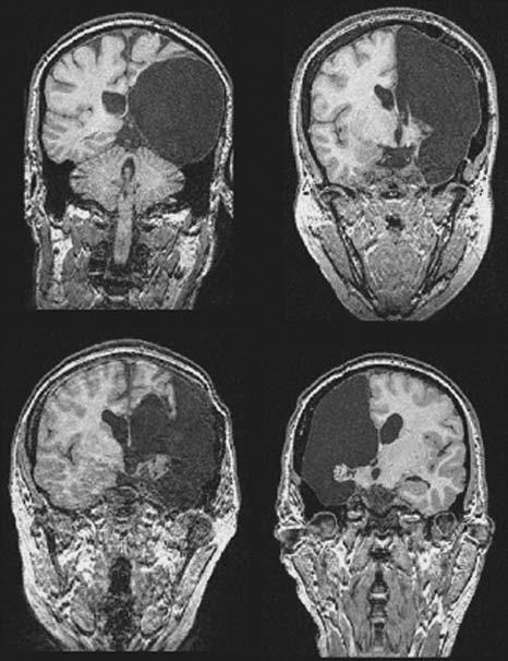
Activation of V5, V3 and V3A visual areas of the intact hemisphere following stimulation of the blind field of a patient who had an hemispherectomy. Source: Bittar et al. Neurimage 1999.
Normal and pathophysiological aging
Aging may be accompanied by a significant decrease in visual abilities. These sensory, but also cognitive, disorders can significantly reduce the autonomy of the individual, requiring support from the family and state at considerable costs. It is therefore important, if not essential, to learn about the basic neural mechanisms that underlie normal and pathophysiological aging of the visual system (and, by extension, of the brain) in order to facilitate the development of appropriate diagnostic tools and therapeutic strategies.
The visual function in healthy elderly people is extensively studied by psycho-physical approaches (including methods of virtual reality) and by functional imaging (optical imaging, EEG, MEG and fMRI): shape, color and movement (especially optic flow, since locomotion is an important factor in autonomy) discrimination, as well as stereo perception are studied. In parallel, we study the visual function of patients with specific pathologies, namely those inducing attention deficits and visual neglect. This work will enable us to develop tools to improve learning and rehabilitation of visual functions.
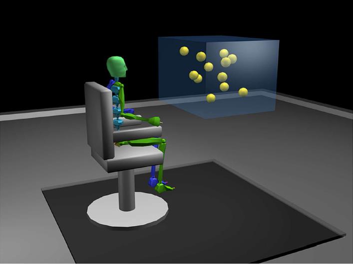
The use of virtual reality to study the visual and attentional abilities in elderly people. Source: Legault, Allard & Faubert. Front Psychol. 2013, 4:323.
Finally, studies are carried out to determine the role of neurotransmitters and neuromodulators in visual function and their impact on the sensory deficits associated with degenerative diseases, such as Parkinson’s and Alzheimer’s disease. Considering the aging of the population, the incidence of neurodegenerative diseases is increasing, and it is essential to determine the mechanisms that lead to the sensory losses observed in the affected patients in order to develop therapeutic tools that would improve their quality of life.
