Research topics of the Cornea and Anterior Segment axis
Corneal wound healing
Corneal wound healing is a major challenge in many pathologies of the ocular surface. In a model using tissue-engineered extracellular matrices, we observed a significant increase in the expression of several genes encoding different subunits of membrane receptors collectively known as integrins, which play an important role in the adhesion and proliferation of corneal epithelial cells. Our new gene profiling platform on DNA microarrays will bring this project to a level never reached before in the study of corneal wound healing. These studies will improve our understanding of the normal and pathological corneal re-epithelialization in order to facilitate the healing of chronic epitheliopathies and to reduce the occurrence of their complications.
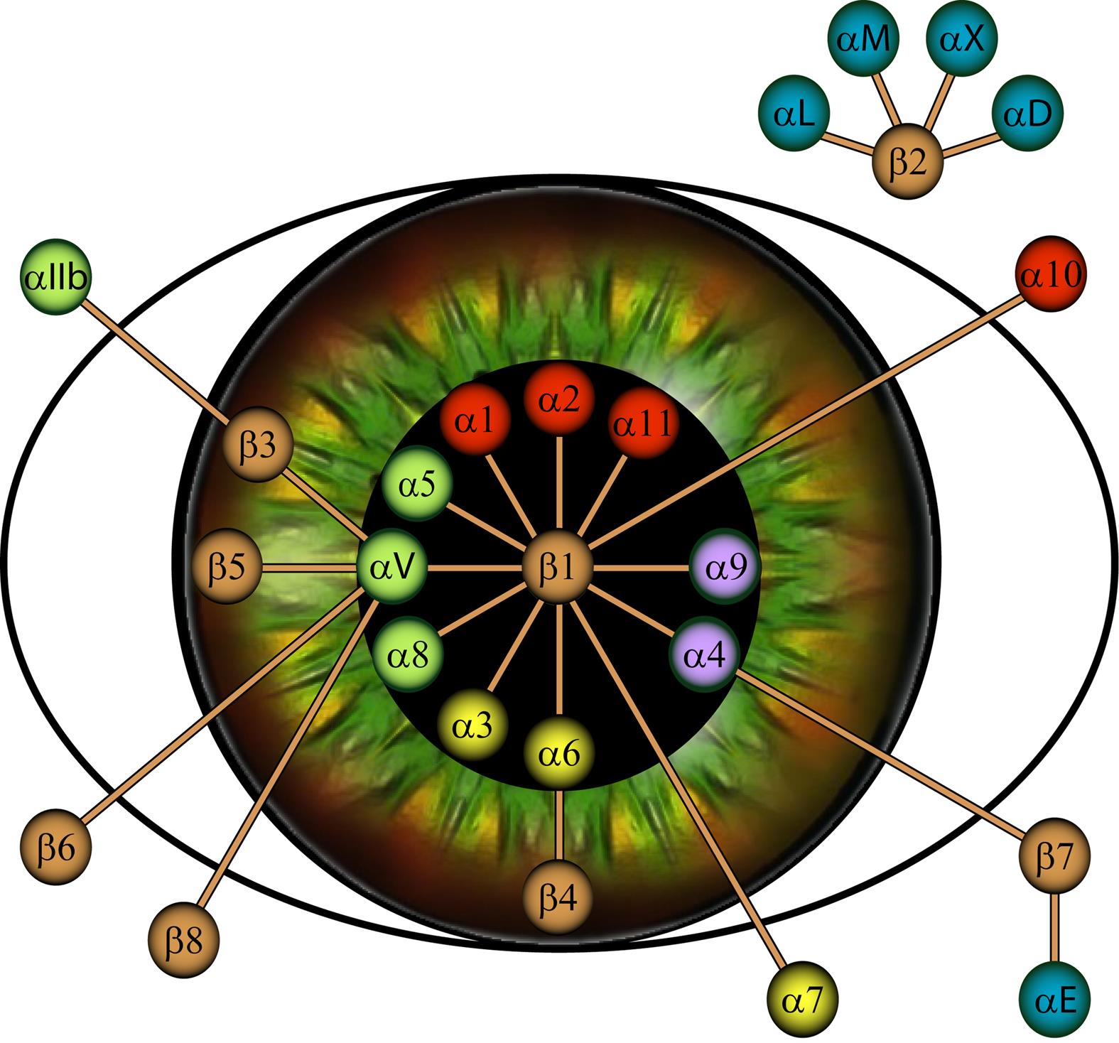
Source : Vigneault, F., Gaudreault, M., Zaniolo, K., Gingras, M.E., and Guérin, S.L. (2007) « Regulation of integrins gene expression in the eye » Progress Retinal Eye Res., 26:99-161.
Toxicity of sun radiation to the human cornea
This research topic has two main components. The first one is to understand the molecular mechanisms protecting the cornea from the genotoxic effects of ultraviolet (UV) radiation. Similar to the skin, the cornea is exposed to UV light, but no cancers linked to this exposure have been reported to date. Thus, our studies seek to understand the mechanisms by which the cornea manages to avoid the malignant transformation induced by UV rays in the skin. In the context of the second component, we work towards the understanding of the consequences of the exposure to UV rays on corneal photoaging. The cornea becomes harder and more opaque with age. Traditionally, this phenomenon has been attributed to chronological aging, but our studies show that exposure to UV rays plays an important role in these events and that the corneal changes with age would rather be attributed to photoaging.
Tissue engineering of the cornea
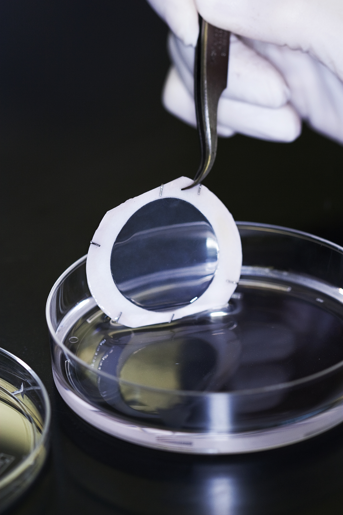
Tissue engineering is an advanced field based on the ability of cells to regenerate tissues and organs. Its two main goals are (i) a better understanding of normal and pathological cellular and molecular mechanisms in the laboratory and (ii) the recovery or replacement of tissues for therapeutic purposes in patients.
- Tissue engineering of the corneal epithelium: These studies aim at the recovery of the pathological epithelial surfaces (chemical burns, neurotrophic degeneration).
- Tissue engineering of the corneal stroma: These studies aim at developing thick and transparent stromal substitutes that respect the anatomical, optical and biomechanical properties of a normal native corneal stroma.
- Tissue engineering of the corneal endothelium: The reconstruction of a corneal endothelium will enable the study of the pathogenesis of age-related endothelial diseases (such as Fuchs’ endothelial dystrophy and pseudophakic keratopathy), as well as the development of replacement tissues to treat these endotheliopathies.
- Tissue engineering of the entire cornea: A full-thickness corneal equivalent will be reconstructed by assembling the three layers: stromal, epithelial and endothelial.
3D analysis of corneal shape
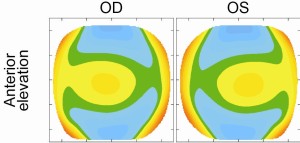
The shape of the cornea is intrinsically linked to its function. The 3D characterization of the shape of the normal and pathological cornea allows a better understanding and treatment of the pathologies affecting its shape. These studies focus on the analysis of the effect of age and other various factors influencing the shape of the normal cornea, such as the degree of ametropia (strength of the glasses) and enantiomorphism (degree of mirror symmetry between the right and left eye). The study of the different stages of Fuchs’ dystrophy progression, as well as the study of wound anatomy and corneal shape before and after penetrating keratoplasty, anterior (DALK) or posterior (DSAEK) lamellar transplants will improve the planification of surgeries involving the different corneal layers. The atlases and image analysis tools used can allow the development of algorithms for the screening and quantification of corneal diseases or refractive surgeries. They also guide the development of corneal substitutes generated by tissue engineering.
3D atlas of the corneal shape. Map of he Anterior elevation. The image on the left illustrates the atlas of the right eye and the image on the right illustrates the left eye.
Laser and corneal surgery
In recent decades, laser technology has led to great leaps in corneal surgery. The research work of several teams of physicists and engineers members of the network deals specifically with the characterization of the corneal ablation parameters by femtosecond lasers and with the development of new high-precision corneal surgical techniques using these lasers. These researchers also work on image analysis algorithms for the characterization of the surface properties of corneal incisions, as well as on unique non-invasive measures of the elasticity and other biomechanical properties of ocular tissues.
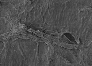
Scanning electron microscopy (SEM) photo photo showing the details of the corneal surface. Marian A, Nada O, Légaré F, Meunier J, Vidal F, Roy S, Brunette I, Costantino S. Smoothness assessment of corneal stromal surfaces. J Cataract Refract Surg. 2013 Jan;39(1):118-27.
Total corneal replacement by PMMA keratoprostheses
The surgical implantation of a PMMA keratoprosthesis (KPro) remains a last resort alternative for end-stage corneal disease too advanced for a corneal transplant. This technique, only recently introduced in Quebec, will be optimized. Cases complicated by dehiscence, infection and glaucoma will be analyzed to improve their understanding and reduce their incidence and severity. The advantages of the improved KPro model for eye anatomy and function will be studied. The impact of this reconstruction by keratoprosthesis on sensory adaptation phenomena and transmodal neuroplasticity following the restoration of visual function will also be studied.
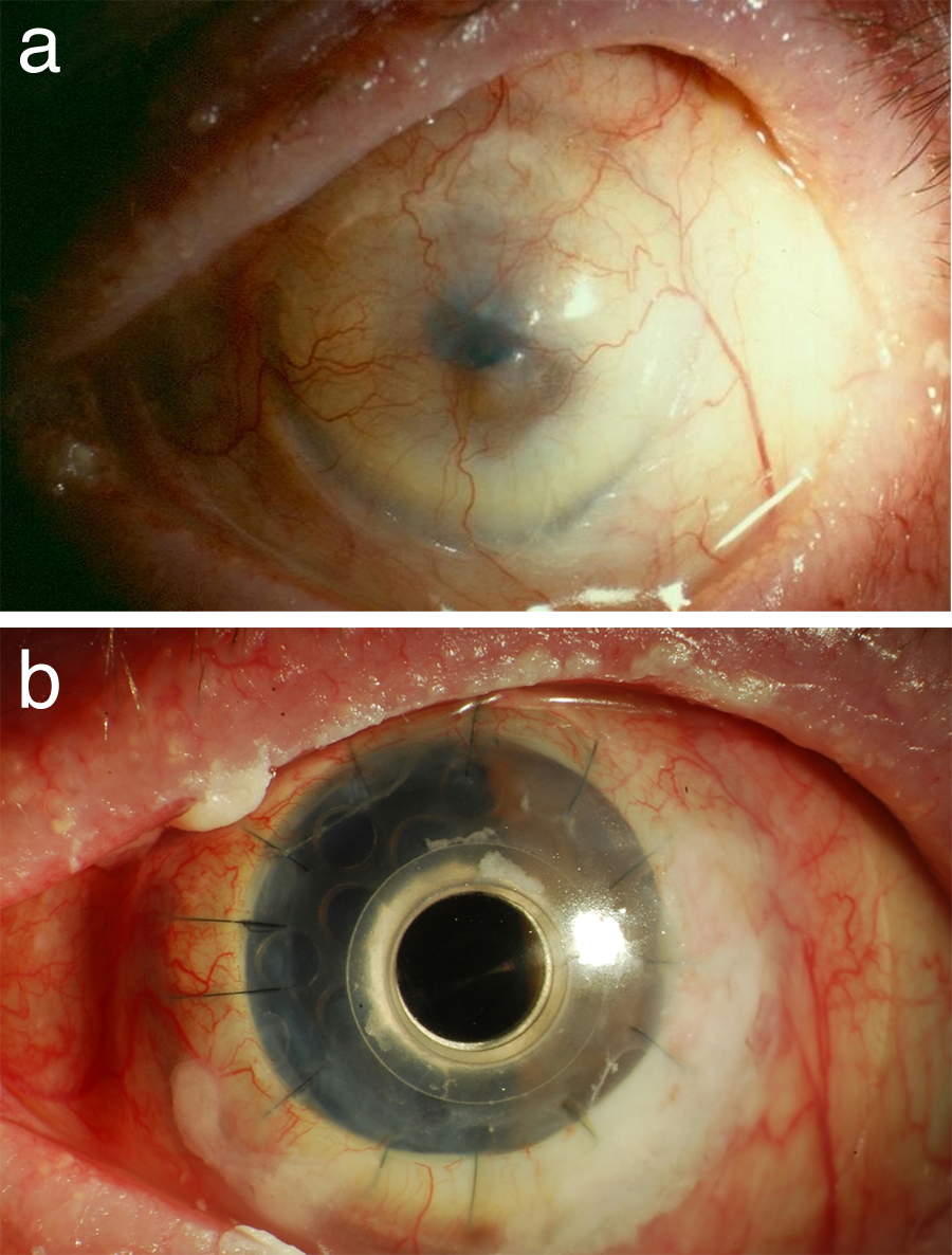
Source: Robert, M.-C. and Harissi-Dagher, M. (2011) « Boston type-1 keratoprosthesis : the CHUM experience. » Can J Ophthalmol. 46(2) :164-8
Regeneration of the trabecular endothelium
Some bone marrow mesenchymal stem cell derived paracrine factors appear to activate progenitor cells in the eye, found namely at the level of the trabecular endothelium. These discoveries open the door to cell therapy for glaucoma.
