

Research topics of the Retina and Posterior Segment axis
The 2013-2017 VHRN scientific program agenda features four major retinal and posterior segment diseases: age-related macular degeneration (AMD), diabetic retinopathy, uveal melanoma and glaucoma.
Age-related macular degeneration
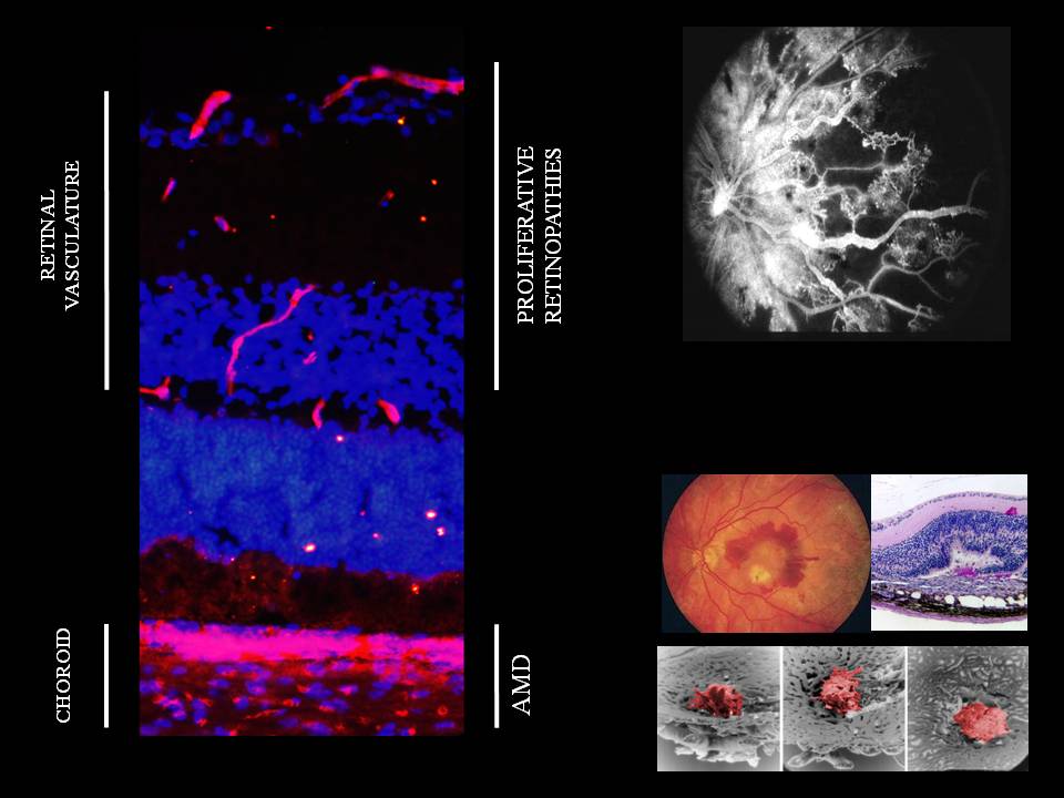
Diabetic retinopathy
Diabetic retinopathy is the third leading cause of blindness in developed countries. It is characterized by the appearance of microaneurysms, vaso-obliteration and retinal oedema, often followed by aberrant pre-retinal neovascularization. Treatments available today only target oedema and neovascularization, without eliminating vascular obliteration and inflammation. Despite the intimate involvement of oxidation and inflammation in the pathogenesis of diabetic retinopathy, the available antioxidants are ineffective, and only steroidal anti-inflammatory drugs have been studied in depth to date. The Retina and Posterior Segment axis researchers are interested in identifying molecules that represent major targets in this disease. Due to their anti-inflammatory properties, mesenchymal stem cells could provide a significant benefit in the treatment of this disease and constitute an area of high-interest in research.
Uveal melanoma
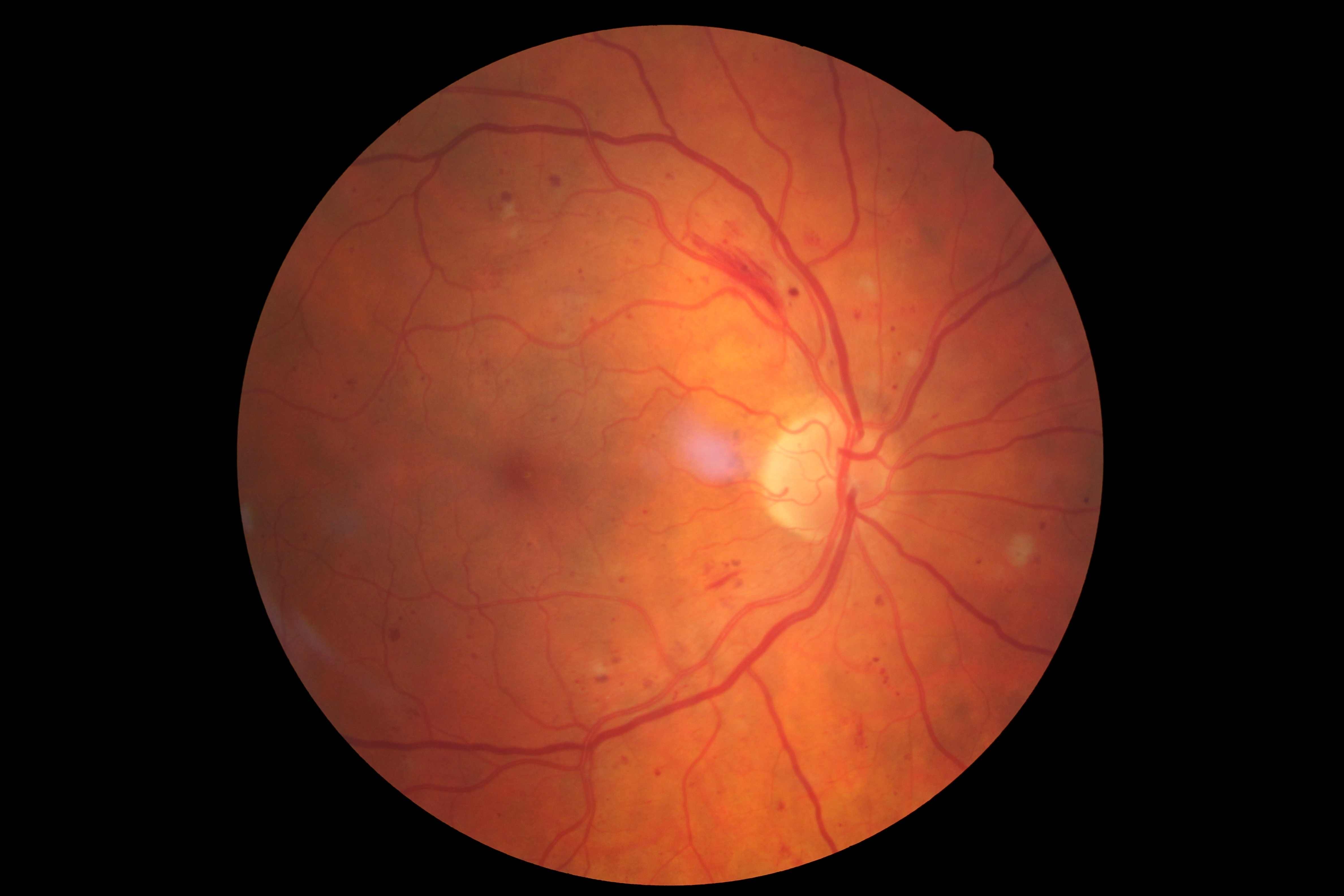
Uveal melanoma is the most common intraocular malignant tumour in adults. This cancer is highly metastatic and notoriously resistant to conventional cancer treatments. Despite an effective treatment of the ocular tumour, more than half of the affected patients die of metastases, mainly hepatic. Micrometastases already present but undetected at the time of the initial diagnosis constitute a major obstacle to successful systemic therapy. The quest for an adjuvant therapy for this malignancy is therefore essential. Several therapeutic avenues are considered by the Retina and Posterior Segment axis researchers. These include the use of specific inhibitors of receptors identified in the molecular signature of tumours with a high metastatic risk, the exploration of the molecular mechanisms involved in the adaptation to hypoxia, and the characterization of the synergistic interactions between cancer cells and stromal cells of the tumoral microenvironment.
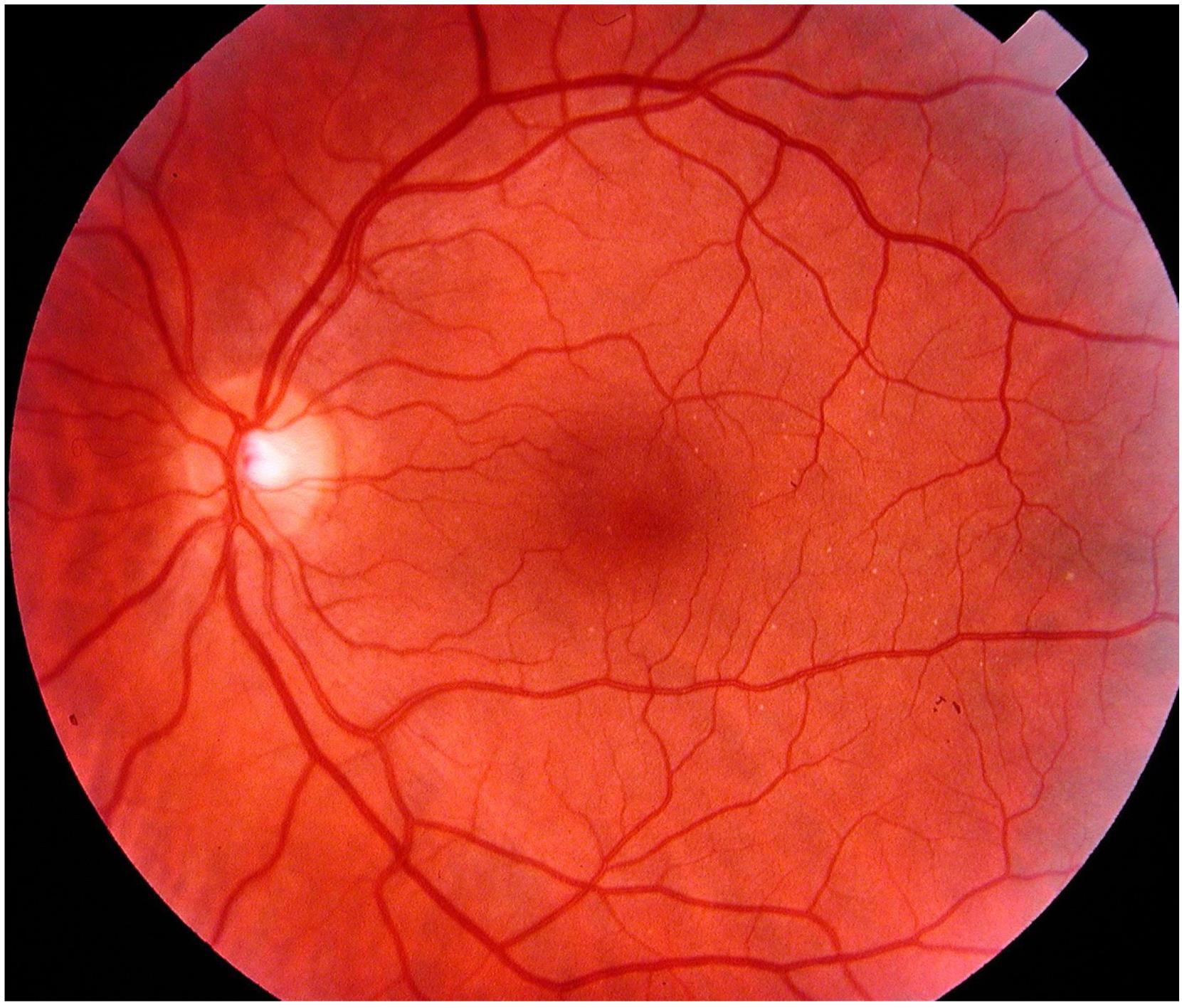
Normal fundus of the eye. The blind spot (white circle on the left) and the blood vessels can clearly be distinguished.
Fundus of the eye in a patient suffering from uveal melanoma. The dark region corresponds to the region where the melanoma is localized.
Glaucoma
Causing retinal and optic nerve neurodegeneration, glaucoma is the leading cause of irreversible but avoidable blindness. It has been estimated that by 2020, over 11 million people will be blind as a consequence of glaucoma worldwide. Increased intraocular pressure (IOP) is considered to be the main risk factor; however, glaucoma can be normotensive. The mechanism by which increased IOP leads to the death of retinal ganglion cells and damages the optic nerve is still poorly understood. The only currently available treatments include ocular hypotensives and surgery. The projects conducted by the various teams of this axis incorporate a multidisciplinary approach based on prevention, early diagnosis and curative and palliative treatment of glaucoma. New pharmacological compounds have been identified and their effectiveness was demonstrated in animals; however, their therapeutic potential for the cortical visual perception still needs to be demonstrated. In addition, a new damaging mechanism based on the stretching of the optic nerve axons has been proposed, as well as a new method of noninvasive imaging.
In glaucoma, the pressure increases inside the eye, which leads to damage to the optic nerve, i.e. to the death of ganglion cells (RGC). In the diagram on the right, the different retinal neuronal and glial cells are depicted; they are grouped into five different layers: ONL, outer nuclear layer, OPL, outer plexiform layer, INL, inner nuclear layer, IPL, inner plexiform layer, GCL, ganglion cell layer. Frédéric Lebrun- Julien from Dr. Adriana Di Polo’s team prepared this image.
Model of the retinal ganglion cell degeneration and regeneration that takes place in glaucoma. From left to right: Transgenic expression of the green fluorescent protein (GFP) in the retinal ganglion cells of adult mice. At higher magnification, immunohistochemical staining of retinal ganglion cells with an anti-tubulin b 3 (red) antibody and after transgenic expression of GFP. Dorsal and ventral view of the left (red) and right (green) ganglion cell axonal projections in the brain. Dendritic tree of an isolated ganglion cell. Fluorescent tracing (red) of regenerating axons in the entire transparent optic nerve of an adult mouse after injury (left). Dr. Vincent Pernet has prepared this image.