Lake J, Zaniolo K, Gaudreault M, Carrier P, Deschambault A, Bazin R, Germain L, Salesse C, Guérin SL. Expression of the α5 integrin gene in corneal epithelial cells cultured on tissue-engineered human extracellular matrices. Biomaterials. 2013 Sep; 34(27):6367-76.
Scientific impact: Corneal reconstruction by tissue engineering is a promising avenue to keep up with the rise in demand for corneal grafts. Proper tissue regeneration requires a good comprehension of the signals that guide this process. This study demonstrated the impact of different components of an extracellular matrix reconstructed by tissue engineering on the expression of the a5 integrin subunit gene from the a5b1 integrin in human corneal epithelial cells. The regulation of a5 by the matrix is of great importance in the healing of corneal wounds as it allows epithelial cells to migrate and adhere properly.
Network contribution: The VHRN (Ocular tissue Bank) has provided the corneal fibroblasts and epithelial cells requires for this project involving many network axes (LG, SLG: cornea and anterior segment; CS: retina and posterior segment; and RB: clinician).
* * *
Original Abstract
Purpose: The integrin α5b1 plays a major role in corneal wound healing by promoting epithelial cell adhesion and migration over the fibronectin matrix secreted as a cellular response to corneal damage. Expression of a5 is induced when rabbit corneal epithelial cells (RCECs) are grown in the presence of fibronectin. Here, we examined whether a5 expression is similarly altered when human corneal epithelial cells (HCECs) are grown on a reconstructed stromal matrix used as an underlying biomaterial. Methods: CAT/a5-promoter recombinant plasmids were transfected into HCECs grown on BSA or on a biomaterial matrix produced by culturing human corneal fibroblasts with ascorbic acid (ECM/35d). The composition of the reconstructed matrix was determined by mass spectrometry and immunofluorescence analyses. Expression of transcription factors (Sp1, AP-1, Pax6 and NFI) that are important regulators of α5 gene expression was monitored by microarrays, qPCR and Western blot. The DNA binding properties of these TFs was determined by EMSAs. Results: Mass spectrometry and immunofluorescence analyses revealed that the biomaterial matrix (ECM/35d) contains several types of collagens, fibronectin, tenascin and proteoglycans. Results from transfection of CAT/a5-promoter plasmids, Western blot, EMSA and microarray analyses indicated that ECM/35d significantly increase expression of the α5 promoter in HCECs as a result of alteration in the expression and DNA binding of the transcription factors NFI, Sp1, AP-1 and Pax6. Furthermore, the ECM/35d increase expression of many MMP genes, most notably that of MMP-9 and MMP-10. Conclusions: The tissue-engineered ECM/35d causes important changes in the activity of the α5 gene promoter as well as genes, such as MMPs, that play important functions in the remodeling of the newly reconstructed stromal matrix. The biological significance of this biomaterial substitute may therefore contribute to better understand the functions played by the a5b1 integrin and MMPs during corneal wound healing.
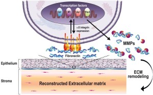
Model of extracellular matrix (ECM) action on the expression of the a5 gene. Through intracellular signalization by their corresponding integrins, the various components from the ECM may alter both the expression and DNA binding properties of the transcription factors (Sp1, NFI, Pax6 and AP-1) that control transcription of the a5 integrin subunit gene, as well as that of multiple genes encoding matrix metalloproteinases (MMPs). The change in the cell surface expression of the a5b1 receptor also translate into corresponding changes in the cell’s adhesive and migratory properties. Meanwhile, the ECM-dependent increased expression of specific MMPs allow an important remodeling of the ECM that favors both of these processes (cell adhesion and migration).


